Radiopacity in Medical Devices
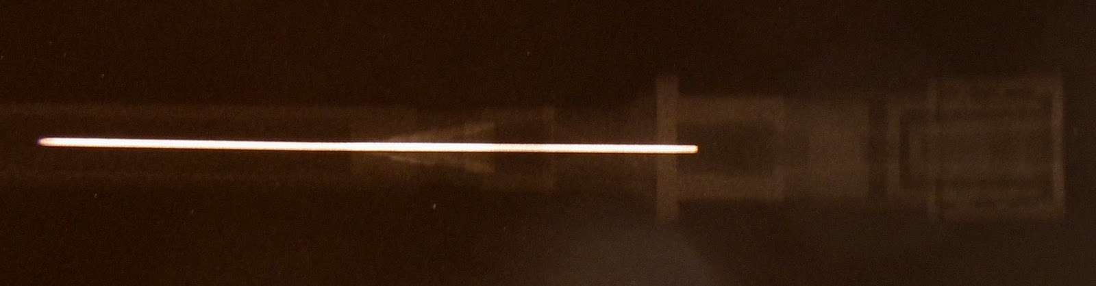
Temporary or permanent implants often contain a radiopacifier, which is a material with a higher electron density contrast compared to the surrounding material so that it absorbs X-ray energy. In an X-ray, a radiopacifier appears as a bright section, as shown in the catheter above (the internal wire is a radiopacifier). Radiopacifiers are often made of metals such as gold, tungston, or powders such as zirconium oxide, barium sulphate and bismuth. When considering the design of a new medical device, manufacturers will need to assess the radiocontrast of the device so that the medical practitioner can see the device during implantation, in the case of catheters, guidewires, and other temporary devices with the use of fluoroscopes, or after permanent implantation, in the case of hip and knee replacements, stents, heart valves, and other permanent devices.
ASTM F640 "Standard Test Methods for Determining the Radiopacity for Medical Use" describes test methods for quantitative assessment of the contrast a radiopacifier has in a medical device, for either permanent implantation or temporary. In this method, the device is placed into an X-ray imaging system and imaged using standard times, voltages, and currents used for the X-ray diagnosis of humans. For two of the test methods, body mimics can be used, which may be animal, cadaver, or synthetic components that replicate the portion of the body where the device is to be placed. From the X-ray image of the device, a densitometry system is used to measure the optical density difference between the sample radiopacifier and the background.
CPG performs ASTM F640 using our custom densitometry system. Please contact us for your testing needs.