Morphological Analysis
Cambridge Polymer Group has extensive experience in visualizing and understanding a diverse range of materials. We have worked with metals and polymers, as well as coated materials and fibers. We have also performed extensive analysis on relatively intractable materials like hydrogels and human tissue. CPG can therefore suggest the most appropriate imaging technique to view structures in and on your materials. We will handle sample preparation, test design, and analysis of the images obtained by a variety of microscopic techniques.
Determine particle size distribution, multi-phase structure, fracture mechanisms, and a host of other applications with microscopy. Cambridge Polymer Group has expertise in a variety of microscopic techniques.
Electron microscopy
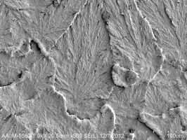
- Scanning Electron Microscopy (SEM): high vacuum, high resolution SEM to evaluate surface structure. Environmental scanning electron microscopy can also be performed on hydrated samples
- Energy Dispersive Spectroscopy (EDS): provides elemental information about the composition of the structure of the surface of a sample. Performed in conjunction with SEM. Elements with atomic numbers down to carbon can be viewed with EDS.
- Transmission Electron Microscopy (TEM): high resolution microscopy showing bulk structure based on electron density differences.
Optical Microscopy
- Transmission, Reflection
- Birefringence
- Hot Stage Microscopy
Particle Size Analysis
- Dynamic Light Scattering
- Malvern Laser
- Optical Microscopy
- Gilsonic Autosiever