
Over the past 50 years, the prevalence of plastics in our consumer waste stream has increased significantly as these materials have become more widely used in products, packaging, and single-use disposable items like water bottles. There has been considerable discussion about the fate of plastics, with coverage often focusing on recycling facilities, landfills, and ocean “garbage patches” in places like the North Atlantic Gyre. More recently, however, attention has shifted toward microplastics as a critical component of plastic pollution, including emerging evidence that these particles can accumulate in human organs such as the liver, kidneys, and brain.
What Exactly Are Micro- and Nanoplastics?
Microplastics are polymer-based particulates with a size range between 0.1 micrometers to 5 mm; while nanoplastics extend from 0.1 micrometers to 1 nm. These particles arise from the breakdown of larger plastic items, as well as from direct sources such as fibers shed from textiles, industrial abrasives, cosmetic additives, and wear debris from polymer components.
Although currently the impact of these materials is largely unknown, toxicologists and clinical researchers are investigating the potential harm of microplastics and nanoplastics on human and animal health. Irrespective of the size of the particles, a key part of this investigation is to identify and quantify the types of polymers making up the particulates, as toxicological studies consider the amount of a particular compound in the body when evaluating the toxicological response (e.g. inflammation, necrosis, etc.), following the old adage “dosis sola facit venenum”, or “the dose makes the poison.” In other words, for a toxic compound to be harmful in the body, it has to reach a threshold in concentration in a biological system.
How Do We Look for Microplastics and Nanoplastics?
Historically, much of the work to detect microplastics in tissues has relied on manual microscopy, sometimes preceded by tissue digestion processes to allow separation of unknown particles. This approach is laborious and faces difficulties in accurately identifying and quantitating the microplastics in the tissue. These difficulties stem from sub-sampling challenges and concerns about the quality and consistency of the recovery process. Due to the resolution limits of microscopy, this approach is unable to see nanoplastics, or even the lower range of microplastics.
These smaller particles are of particular interest because a large fraction of the material may exist below conventional optical resolution limits. Although the total volume of these particles is not substantial, this population may be significant in terms of the number of particles. Capturing this population is important, because absolute dose is only part of the concern, and the surface area-to-volume ratio may also be significant. Smaller particles present more surface area per unit mass, which can influence reactivity and biological interactions. One can see that intuitively because a medical implant made from a polymer has been validated to be safe, but it is not clear whether nanoplastics of the same polymer, capable of migrating to different organs, should be considered safe.
The biological impact of micro- and nano-plastics are not yet well understood, but having analytical tools to identify and quantify them is an essential step toward determining if they contribute to clinical issues.
Py-GC/MS: Looking Inside Tissues without Seeing the Particles
Consequently, researchers are turning to other techniques to identify and quantitate these smaller particles. An emerging technique is pyrolysis gas chromatography with mass spectroscopy (Py-GC/MS), which allows analysis of particulates (both micro and nano) in their native tissue matrices. In Py-GC/MS, the sample is thermally decomposed, and the resulting fragments are separated and analyzed to generate polymer-specific fingerprints.
This approach offers several advantages for micro- and nanoplastic analysis:
- Direct analysis of intact tissues or partially processed matrices.
- Polymer-specific identification and quantitation, even when particles are below optical resolution.
- Simultaneous detection of multiple common plastic types.
Using Py-GC/MS, it is possible to distinguish and measure polymers such as polyurethanes, polymethyl methacrylates, polystyrenes, polyvinyl chloride, polyethylene, polypropylene, nylons, polyesters, and polycarbonates in biological samples.
Cambridge Polymer Group has recently developed assays using Py-GC/MS to investigate micro and nano-plastics in lung tissue from certain high-risk industries, providing a path to more robust exposure assessment and materials for subsequent toxicological evaluation.
What Is Being Found in Human Organs?
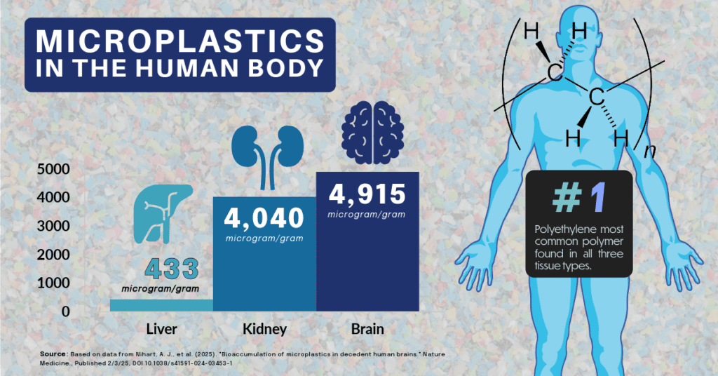
A recent study published by Nihart and colleagues (2025)[1] used Py-GC/MS to examine micro and nanoplastics in human livers, kidneys and brains. They obtained tissues from autopsy specimens from 2015-2024.
Their analysis showed consistent levels of plastic particulates in the liver and kidney specimens at concentration levels of 433 and 4,040 microgram/g, respectively. The plastic levels in the brain were much higher, reaching median levels of 4,915 microgram/g in the 2024 samples. The authors note that brain tissue from patients diagnosed with dementia had a notably higher concentration of particulates.
Interestingly, polyethylene was the most common type of polymer found in all three tissue types, particularly for the brain tissue. No hypothesis is offered by the authors as to why this polymer may be more prevalent.
Polyethylene is a commonly used commercial polymer with many applications ranging from supermarket bags to water bottles. Arguably one would expect to see more polyesters and polyvinyl chlorides, given their abundance in single-use packaging systems. One potential source of polyethylene particulates is from hip and knee replacements, where the bearing surface is often comprised of a form of polyethylene, and if this bearing surface undergoes wear, polyethylene particles are generated. The study did not examine this potential source. If these particles are indeed from orthopedic implants the data suggests that particles can migrate throughout the body, irrespective of source. A future study may attempt to correlate observed polyethylene particulates with the presence of a hip or knee replacement in the patient. As polyethylenes used in these implants have improved in the past 20 years, we would expect to see debris from these types of implants to decrease.
Where Does This Leave Materials and Medical Device Developers?
For stakeholders in materials science, medical devices, and occupational health, these findings carry several implications:
- Material selection and wear performance: Understanding how wear particles distribute beyond the implantation site may influence material choices and design strategies for implants and other long-term devices.
- Exposure assessment in high-risk settings: Industries involving fine plastic particulates, thermal processing, or high-energy machining may benefit from targeted tissue or biological fluid screening for micro- and nanoplastics.
- Regulatory and toxicological frameworks: As tools like Py-GC/MS make it possible to characterize internal plastic burdens more accurately, regulatory expectations and risk assessment paradigms are likely to evolve.
- Exposure appears to be inevitable: Although the Nihart study is for a relatively small population, the data suggests that no one is immune to this risk, and consideration of the reduction of single use applications and more efficient recycling is clearly going to be of importance.
Cambridge Polymer Group supports this emerging area with Py-GC/MS-based micro- and nanoplastic assays for biological and environmental matrices, as well as complementary materials characterization services. If you are evaluating plastic exposure in a high-risk population, characterizing wear debris from a new device, or designing a toxicology study that requires polymer-specific quantitation, our team can help you develop an appropriate analytical strategy.
To discuss a specific application or study design, please contact us to speak with one of our scientists.
[1] Nihart, A. J., et al. (2025). “Bioaccumulation of microplastics in decedent human brains.” Nature Medicine., Published 2/3/25, DOI 10.1038/s41591-024-03453-1