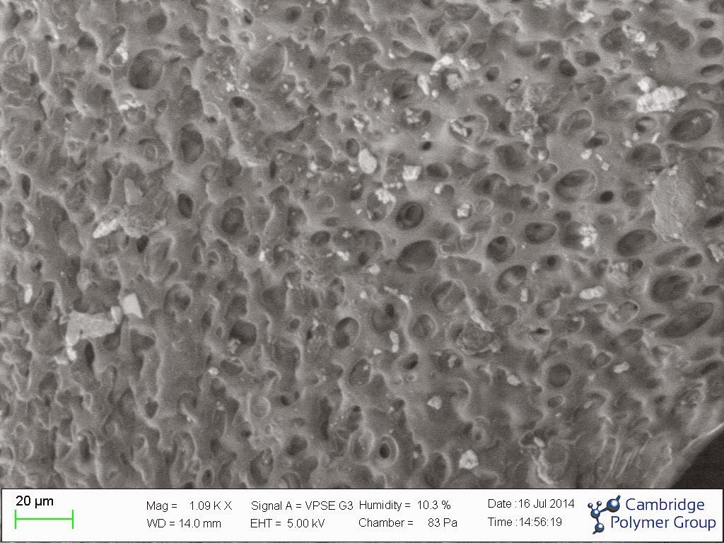
Variable Pressure Scanning Electron Microscopy

Scanning electron microscopy (SEM) is a powerful imaging technique that can be used to discern morphological features down to nanometers. High energy electrons are focused into a narrow beam with electro-magnets, which then impinge on the sample and scatter backward off the surface. This beam is rastered across the surface of the sample in a similar manner to old television cathode ray tubes. Detectors then create an image by collecting these scattered electrons at each raster point, either from the source electrons that are backscattered from the surface, or from secondary electrons that are stripped from the atoms by the source electrons on and below the surface of the sample. Because of the flood of electrons (essentially a current) on the surface of the samples, a conductive path is required to prevent charge buildup. Charge buildup will alter the appearance of the surface by deflecting the paths of the electrons. Normally non-conductive materials such as polymers and other organic species such as tissue must be sputter-coated with a conductive coating, such as gold or carbon, in order to prevent this charge buildup. This process will hence alter the surface properties of the sample. Additionally, high vacuum is required with this approach, which will dehydrate samples containing a volatile material such as water. The surface coating will also interfere with energy dispersive spectroscopic analysis of the surface of the sample (EDS), whereby the metallized coating may obscure the elements actually present in the sample.
As an alternative method, variable pressure SEM, which is also sometimes called environmental SEM, can be used. Here a controlled amount of gas, which can be inert, such as nitrogen, or contain water vapor, is maintained in the sample chamber, while an aperture inhibits the flow of this gas into the gun chamber. The gas in the chamber is ionized by the incoming source electrons, and hence will neutralize charge buildup on the sample, negating the need for gold coating. Additionally, the increased gas pressure slows the evaporation of liquids, allowing the visualization of water-containing samples in a hydrated state, all be it at a reduced resolution.
CPG recently acquired a variable pressure scanning electron microscope (SEM). This system is useful for failure analysis of components, in that the imaging is non-destructive. Additionally, the CPG system has a cold stage, which allows the control of relative humidity and temperature in the chamber, which is useful for imaging water-containing samples such as tissue and hydrogels. The system acquired also has a large capacity chamber enabling in most cases visualization on entire devices and components.
More information can be found here.