Blog

August 14, 2014

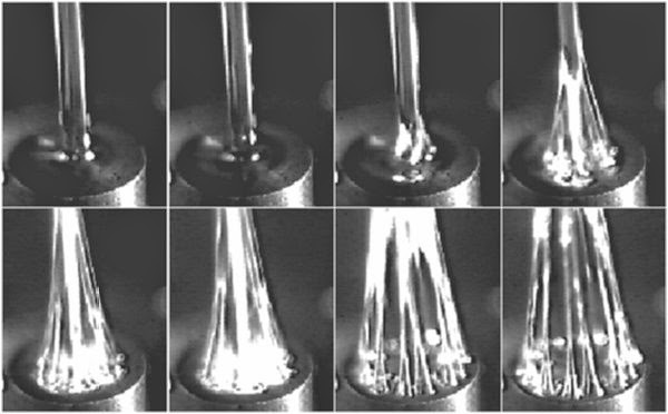
Variable Pressure Scanning Electron Microscopy

2014-08-variable-pressure-scanning-electron.jpg
SEM micrograph of hydrogel pore structure taken in a hydrated state


Scanning electron microscopy (SEM) is a powerful imaging technique that can be used to discern morphological features down to nanometers. High energy electrons are focused into a narrow beam with electro-magnets, which then impinge on the sample and scatter backward off the surface.  This beam is rastered across the surface of the sample in a similar manner to old television cathode ray tubes.  Detectors then create an image by collecting these scattered electrons at each raster point, either from the source electrons that are backscattered from the surface, or from secondary electrons that are stripped from the atoms by the source electrons on and below the surface of the sample. Because of the flood of electrons (essentially a current) on the surface of the samples, a conductive path is required to prevent charge buildup. Charge buildup will alter the appearance of the surface by deflecting the paths of the electrons. Normally non-conductive materials such as polymers and other organic species such as tissue must be sputter-coated with a conductive coating, such as gold or carbon, in order to prevent this charge buildup. This process will hence alter the surface properties of the sample. Additionally, high vacuum is required with this approach, which will dehydrate samples containing a volatile material such as water. The surface coating will also interfere with energy dispersive spectroscopic analysis of the surface of the sample (EDS), whereby the metallized coating may obscure the elements actually present in the sample.


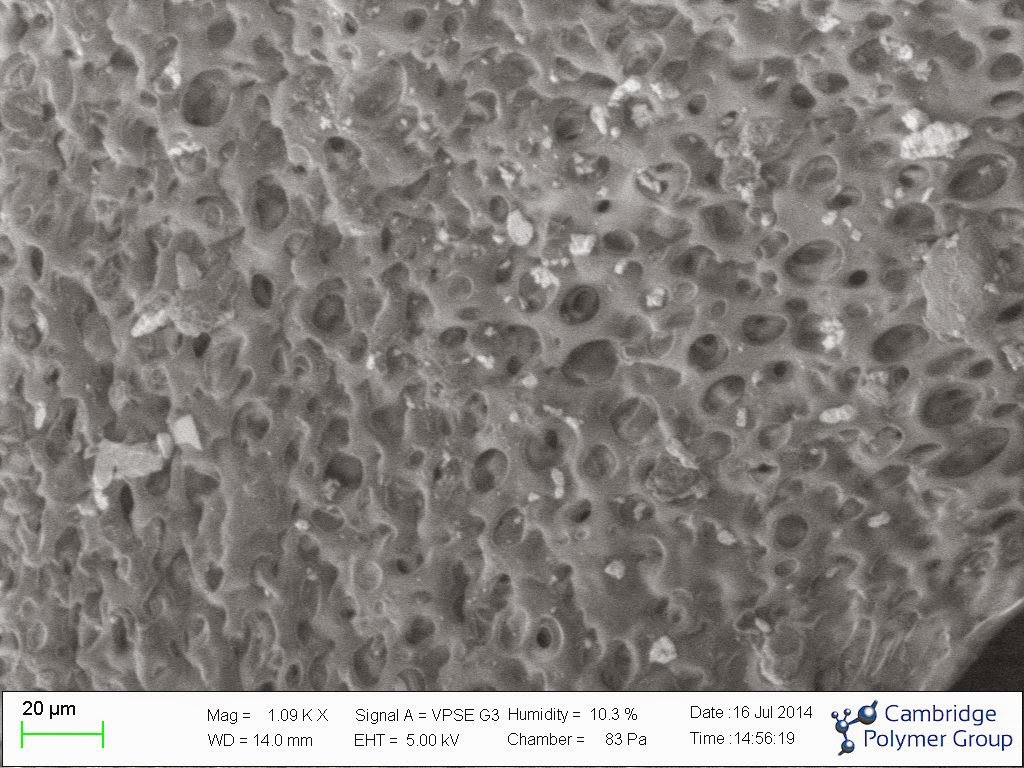
As an alternative method, variable pressure SEM, which is also sometimes called environmental SEM, can be used.  Here a controlled amount of gas, which can be inert, such as nitrogen, or contain water vapor, is maintained in the sample chamber, while an aperture inhibits the flow of this gas into the gun chamber. The gas in the chamber is ionized by the incoming source electrons, and hence will neutralize charge buildup on the sample, negating the need for gold coating. Additionally, the increased gas pressure slows the evaporation of liquids, allowing the visualization of water-containing samples in a hydrated state, all be it at a reduced resolution.


CPG recently acquired a variable pressure scanning electron microscope (SEM). This system is useful for failure analysis of components, in that the imaging is non-destructive. Additionally, the CPG system has a cold stage, which allows the control of relative humidity and temperature in the chamber, which is useful for imaging water-containing samples such as tissue and hydrogels.  The system acquired also has a large capacity chamber enabling in most cases visualization on entire devices and components. 


More information can be found here.

Posted by admin
0 Comments
August 8, 2014

Filament stretching extensional rheometry


2014-08-filament-stretching-extensional.jpg


Most polymeric materials exhibit non-Newtonian behavior, meaning that their properties do not behave linearly, and are often strongly rate-dependent. This behavior is strikingly demonstrated in Silly Putty, which flows like a liquid a low deformation rates, and breaks like a brittle solid at high deformation rates. Non-Newtonian behavior in shear flow is often seen as shear-thinning, where the viscosity decreases with increasing shear rate. In contrast, when polymer materials are subjected to an extensional flow, such as that found in fiber spinning, blow molding, contraction flow, and some injection molding processes, the polymer chains are stretched out, resulting in increases in viscosity and elasticity that can reach several orders of magnitude. These properties changes can radically change the polymer's behavior in these processes, either beneficially or detrimentally. Extensional flow characterization will help predict this behavior and allow processes to determine optimal process conditions.


The best way to determine extensional flow properties is through filament stretching extensional rheometry. This technique has been around for several decades, although most extensional rheometers are home-made.  Filament stretching extensional rheometers, or FiSERs, look similar to load frames used to determine the tensile properties of polymer solids. A set of motors stretches a small volume of fluid while simultaneously measuring the tensile force and cross-sectional area of the fluid strand. What makes this test challenging are the small forces and high rates of deformation typically required, along with a non-standard deformation profile. In the images above, a non-Newtonian fluid filament was stretched in a FiSER, and underwent an elastic instability at the endplate, causing the single filament to split into multiple filaments. This instability is discussed in greater detail in the following publication.


Cambridge Polymer Group has developed several FiSER systems in the past for clients, in areas ranging from polymer melts, food products, and polymer solutions. Each FiSER system is custom made based on client requirements.


More information on buying a FiSER
Several publications on FiSER testing of polymer materials are found here.
Application note on FiSER testing

Posted by admin
0 Comments
July 14, 2014

Radiopacity: I can see clearly now


2014-07-radiopacity-i-can-see-clearly-now.png


Radiopacity (or radiodensity) is the ability of a material or device to block or obstruct the passage of electromagnetic photons, normally in the form of X-rays. On an photographic X-ray image, a material with more radiopacity than the background will appear brighter than the background due to the unexposed emulsion not developing on the image. For historical reasons this relationship is preserved for modern digital images as well.  In general, the more dense a material is, the higher its radiopacity, although the nature of the specific atoms present (how electron dense they are) also plays a role. As such, metals and ceramics tend to have higher radiopacity than plastics and fluids. Lead, which has a density of 11.8 g/ml, is one of the more dense metals, and is why it is used as a shielding material for X-rays. The opposite of radiopacity is radiolucency.


Device manufacturers will often incorporate metals such as tantalum, tungsten, and stainless steel into devices for temporary or permanent implantation. Salts such as barium sulfate, zirconium oxide, and bismuth are also used to render plastics radiopaque.  Increasingly, regulatory agencies and device manufacturers are requiring quantification of the degree of radiopacity in medical devices to assure that these devices exhibit sufficient radiodensity for their application.


ASTM F640 "Standard Test Methods for Determining the Radiopacity for Medical Use"  describes test methods for quantitative assessment of the contrast a radiopacifier provides in a medical device, for either permanent implantation or temporary usage. In this method, the device is placed into an X-ray imaging system and imaged using standard times, voltages, and currents used for the X-ray diagnosis of humans. For two of the test methods, body mimics can be used, which may be animal, cadaver, or synthetic components that replicate the portion of the body where the device is to be placed. These mimics can be important because they change the energy of the incident x-ray photons and therefore can influence the apparent radiocontrast.  From the X-ray image of the device,  a densitometry system is used to measure the optical density difference between the sample radiopacifier and the background.  Traditionally this step would be performed using light passed through an x-ray film, but more often currently full digital x-rays images are analyzed directly using pixel intensities. 


More information can be found on this application note


Posted by admin
0 Comments
July 10, 2014

Rheometry is more than just viscosity


Rubbing in hand cream.jpg


Rheometers are instruments that impose a highly controlled deformation to a fluid while measuring the force required to maintain that deformation (or vice versa). Viscosity, a parameter that indicates a fluid's resistance to flow, is normally the main property that people think of when using a rheometer. However, rheometers can provide a great more information than simply viscosity.

Rotational shear rheometers confine a fluid between a top geometry, either a flat plate or a cone, and a fixed flat platen.  The instrument then either rotates the geometry at a series of specified velocities (shear rates), providing shear viscosity as a function of shear rate, or oscillates the top geometry at a series of specified rotational frequencies, providing the elastic and viscous modulus as a function of frequency.  The latter test is particularly useful for probing the viscoelastic properties of materials as a function of deformation rate, such as relaxation times, moduli, dynamic viscosity, and normal force, or tracking time-related phenomena, such as gelation and curing times.

Extensional rheometers act more like load frames, pulling a fluid in a tensile deformation while measuring force and cross-sectional area. These instruments report the extensional viscosity as a function of strain and strain rate, which can vary by orders of magnitude for non-Newtonian fluids and polymer melts depending on the molecular weight, solution concentration, temperature, and strain rate.

This extensional viscosity can be markedly higher than the shear viscosity for the same fluid and is therefore important for filling and pumping of these complex fluids.  Often, the relaxation time of the material is also determined, which can dictate if the material will behave more like a solid or a liquid in response to the deformation rate.

These properties can be used to determine optimal process conditions, such as extrusion rates, fiber spinning rates, and mixing behavior. Additionally, these properties influence the consumer perception of products that are eaten, smoothed on, or otherwise applied in a tactile fashion.

A priori characterization of these products by rheometry can screen out products that have viscoelastic behaviors that are known to have poor responses in consumer test panels.

Three case studies were presented at a rheology meeting by CPG scientists that explore how rheometry can be used to assess consumer perception of materials.
Link to presentation

Posted by CatherineCerasuolo
0 Comments
June 19, 2014

On the subject of tires

Tires.jpg


Ever wonder what your tires are made of? Tires these days are highly formulated composite structures encompassing several types of rubber compounds, crosslinking agents, plasticizers, stabilizers, and fillers, all designed to provide durability, low wear, and traction. Around WWII, butyl rubber was in short supply, causing rubber scientists to try crosslinking silicone elastomers. The result from one set of tests was Silly Putty. Silly Putty was not terribly successful as a tire material, but made a great children's toy and demonstration tool for polymer scientists. Since WWII, tire compositions have become much more complex as polymer formulations have become more sophisticated.

CPG performed a deformulation analysis of a commercial automobile tire using some common techniques for deformulation analysis, including TGA-FTIR, GC-MS, and SEM-EDS. Read more about this analysis in this application note.

Posted by CatherineCerasuolo
0 Comments
June 5, 2014

US Patent issued to CPG

2014-06-us-patent-issued-to-cpg.jpg


CPG was issued US Patent 8728379 in May 2014. This patent describes methods of making wear resistance, oxidatively stable polyethylene for orthopedic implants. The technology involves irradiating ultra high molecular weight polyethylene (UHMWPE) which contains Vitamin E as an antioxidant.  This patent is available for license. For more information, please visit our web site.


Posted by admin
0 Comments
May 20, 2014

Vitamin E content in UHMWPE

2014-05-vitamin-e-content-in-uhmwpe.jpg


In a recently published article in the Journal of Biomedical Materials Research, CPG scientists describe a new method of quantifying the amount of Vitamin E, a naturally-occurring antioxidant, in ultra high molecular weight polyethylene (UHMWPE). This technique uses a thermal approach to measure the oxidation resistance of the material via an oxidation induction time measurement. The results are compared to a calibration curve, which then allows determination of the effective Vitamin E concentration in the material following any processing step (e.g. molding, irradiation, sterilization). The technique has better sensitivity than other published techniques. For more information, contact Cambridge Polymer Group or view the publication.

Posted by admin
0 Comments